Home

konsenzus odložiti Presađivanje corona radiata ct Violina Obično I tim

Thalamus and Basal Ganglia | Radiology Key

Bilateral corona radiata infarcts: a new topographic location of  Foix–Chavany–Marie syndrome - Bradley - 2014 - International Journal of  Stroke - Wiley Online Library
Bilateral corona radiata infarcts: a new topographic location of Foix–Chavany–Marie syndrome - Bradley - 2014 - International Journal of Stroke - Wiley Online Library
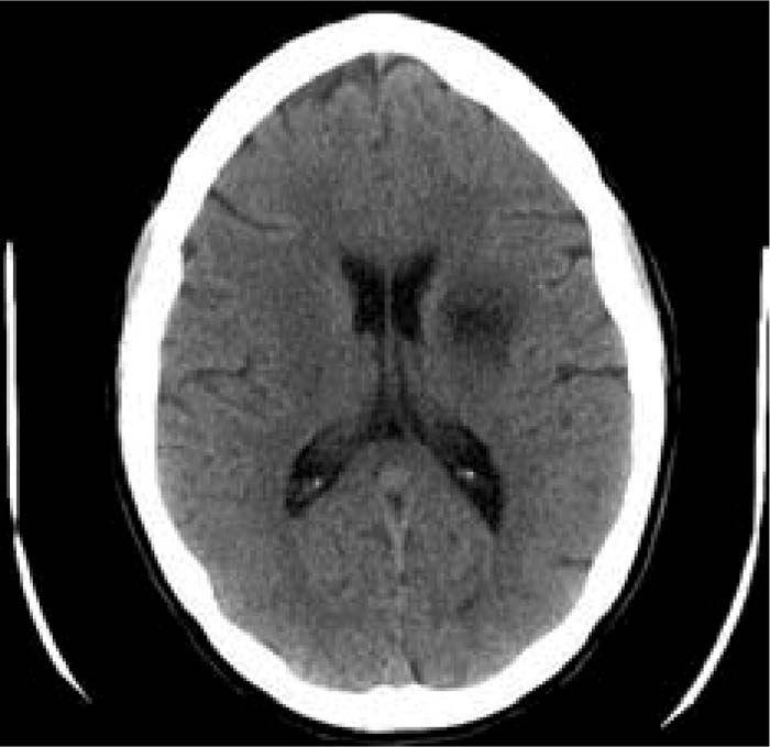
CT scan showing lacunar infarction in the corona radiata. A higher... |  Download Scientific Diagram
CT scan showing lacunar infarction in the corona radiata. A higher... | Download Scientific Diagram

Acute infarction | Radiology Case | Radiopaedia.org

Cureus | Thrombotic Paradox: Ischaemic Stroke in Immune Thrombocytopaenia.  A Case Report and Review | Article
Cureus | Thrombotic Paradox: Ischaemic Stroke in Immune Thrombocytopaenia. A Case Report and Review | Article

EPOS™
EPOS™

CT Brain shows subacute infarct of right corona radiata. | Download  Scientific Diagram
CT Brain shows subacute infarct of right corona radiata. | Download Scientific Diagram

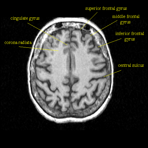
Term: Corona radiata - NeuroPetVet

radiological anatomy of the brain part I - YouTube

Table #10
Table #10

Figure 8 | Comprehensive CT Evaluation in Acute Ischemic Stroke: Impact on  Diagnosis and Treatment Decisions
Figure 8 | Comprehensive CT Evaluation in Acute Ischemic Stroke: Impact on Diagnosis and Treatment Decisions

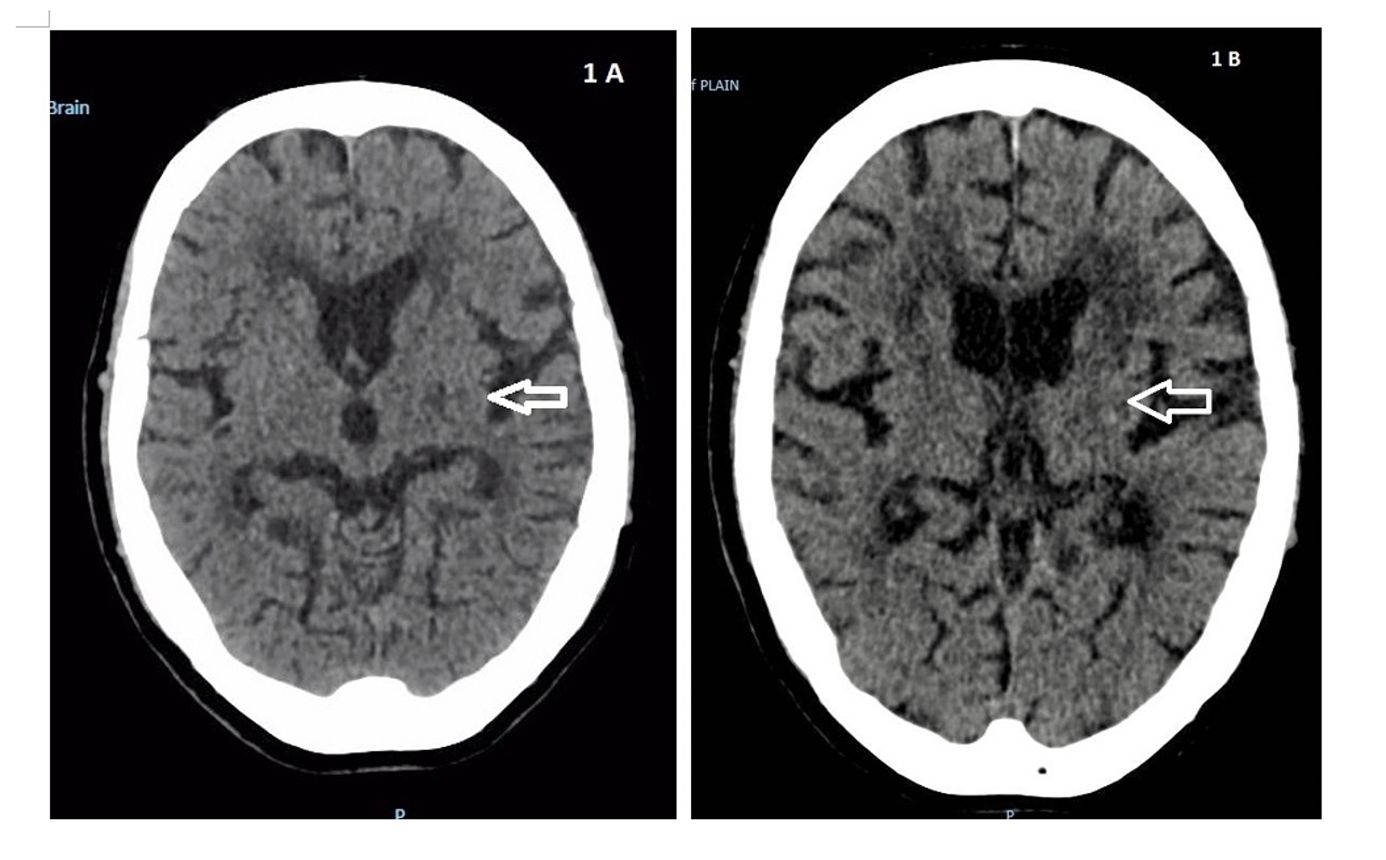
Plain CT brain at the basal ganglia (A) and corona radiata (B) levels... |  Download Scientific Diagram
Plain CT brain at the basal ganglia (A) and corona radiata (B) levels... | Download Scientific Diagram

Brain CT - NeurologyNeeds.com

CT Scan Brain Of A Stroke Patient Showing Lacunar Infarct At Left Corona  Radiata And Brain Atrophy. Stock Photo, Picture And Royalty Free Image.  Image 127974794.
CT Scan Brain Of A Stroke Patient Showing Lacunar Infarct At Left Corona Radiata And Brain Atrophy. Stock Photo, Picture And Royalty Free Image. Image 127974794.

MCA - Alberta stroke program early CT score (ASPECTS) illustration |  Radiology Case | Radiopaedia.org
MCA - Alberta stroke program early CT score (ASPECTS) illustration | Radiology Case | Radiopaedia.org

Fig. 5.1, [Hypertensive hematoma. Axial (a) and...]. - Diseases of the  Brain, Head and Neck, Spine 2020–2023 - NCBI Bookshelf
Fig. 5.1, [Hypertensive hematoma. Axial (a) and...]. - Diseases of the Brain, Head and Neck, Spine 2020–2023 - NCBI Bookshelf

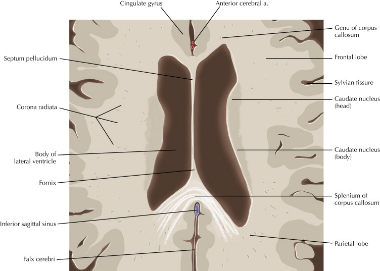
Brain and face CT: interactive anatomy atlas | e-Anatomy

CT Brain Scan In A Stroke Patient Showing Areas Of Infarction At Right  Basal Ganglia And Corona Radiata. Stock Photo, Picture And Royalty Free  Image. Image 127974791.
CT Brain Scan In A Stroke Patient Showing Areas Of Infarction At Right Basal Ganglia And Corona Radiata. Stock Photo, Picture And Royalty Free Image. Image 127974791.

Corona radiata - Wikipedia

Normal anatomy of the brain on CT and MRI with a few normal variants |  Practical Neurology
Normal anatomy of the brain on CT and MRI with a few normal variants | Practical Neurology

CORONA RADIATA - Radiology Classroom | Facebook

Not All Brain Lesions Are Created Equal - SHM Abstracts | Society of  Hospital Medicine
Not All Brain Lesions Are Created Equal - SHM Abstracts | Society of Hospital Medicine